Histology and Analysis


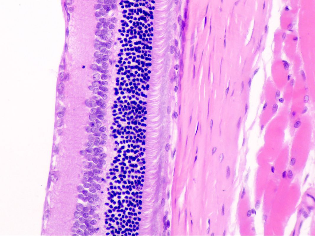 Retina; H&E stain
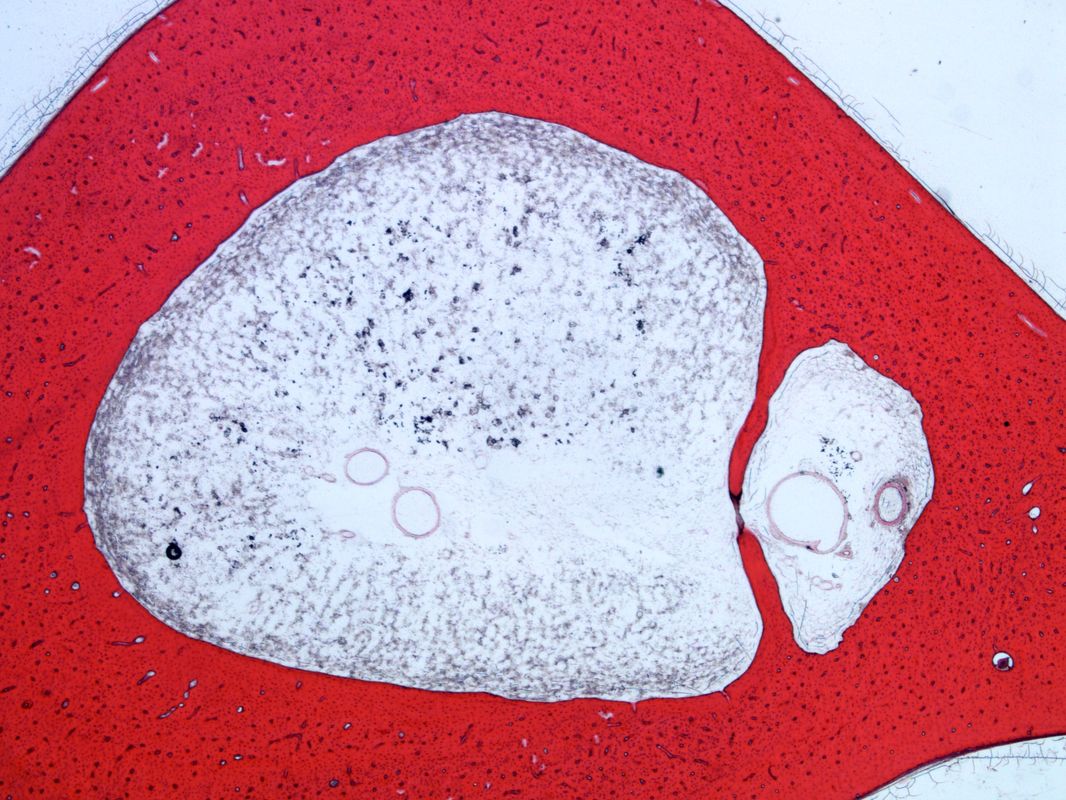
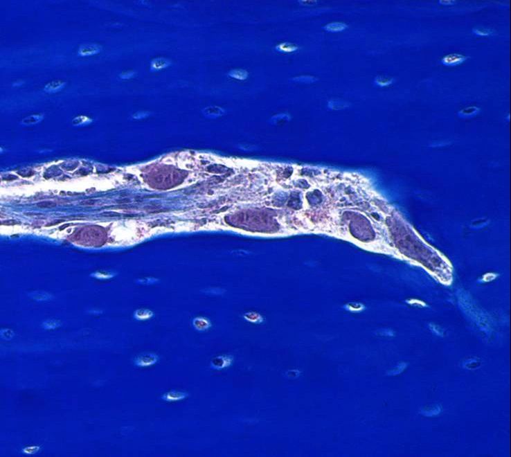
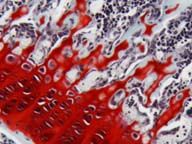
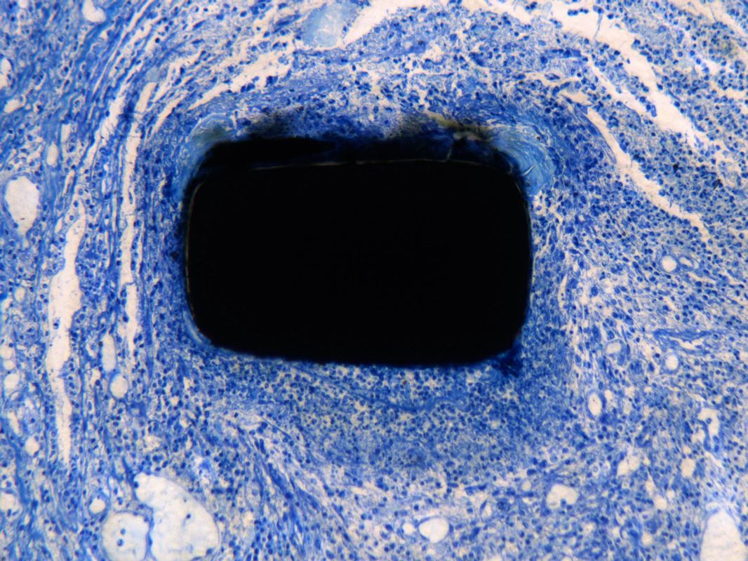 Subcutaneous tissue; Metal implant; Toluidine blue stain
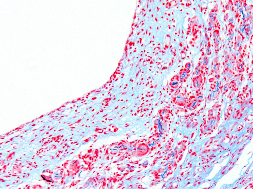
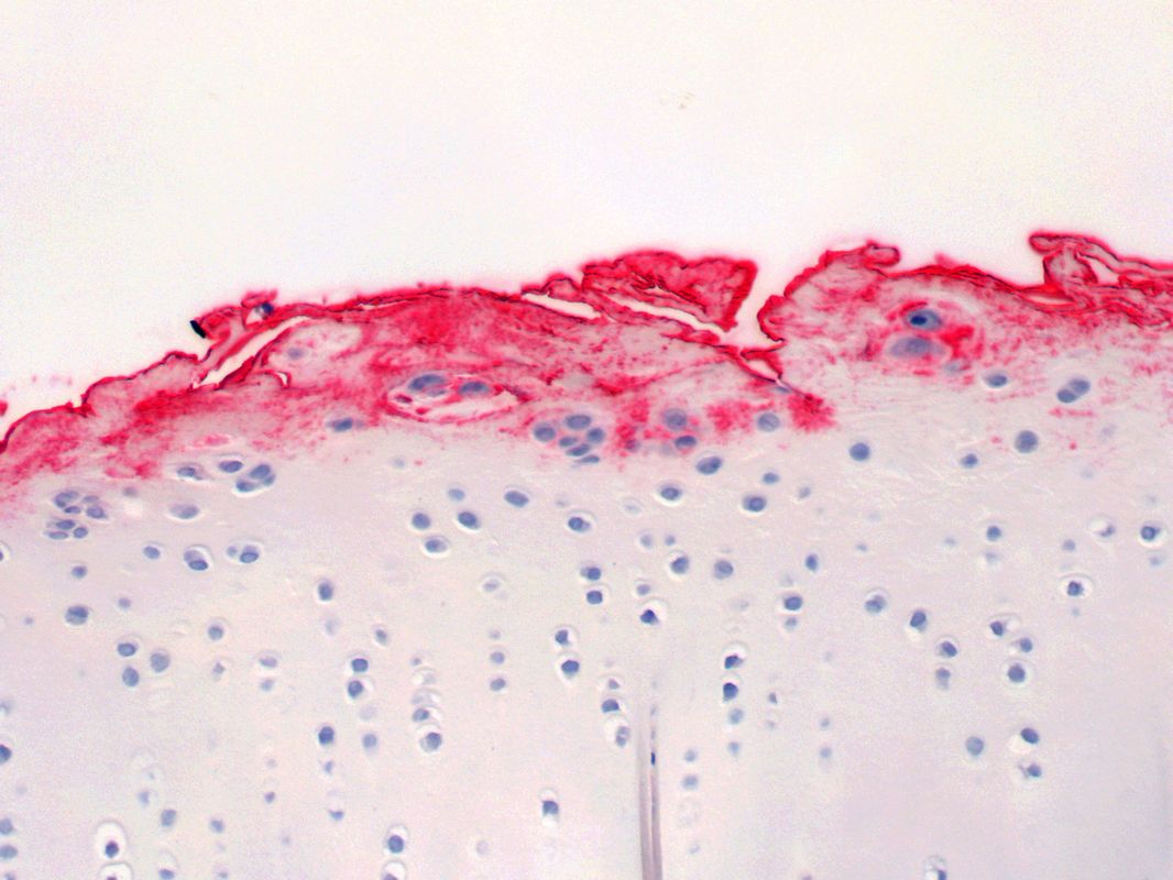
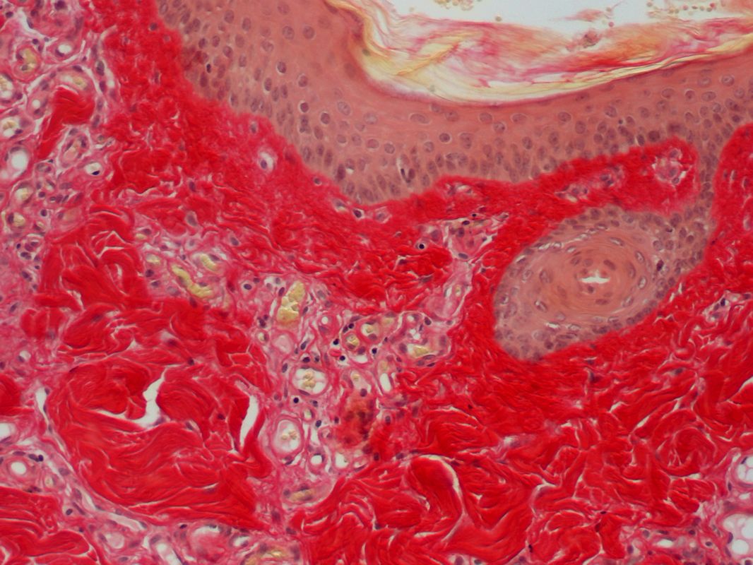 Skin; Picrosirius red stain
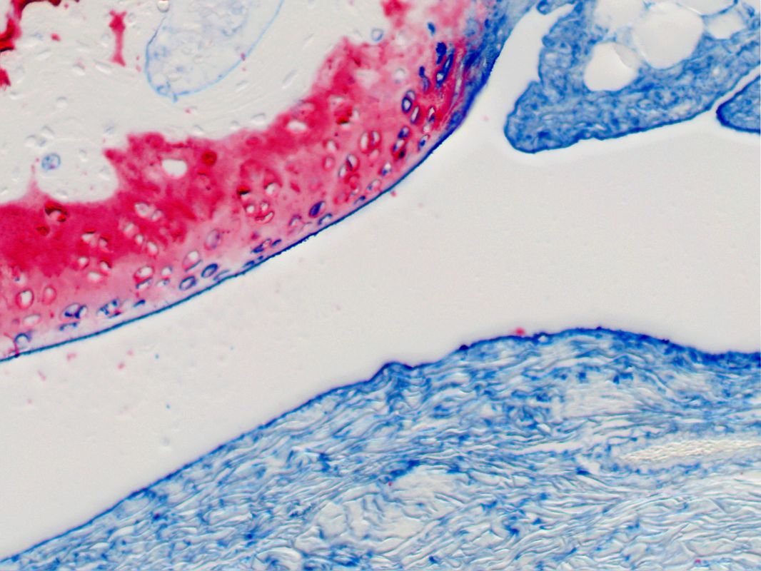
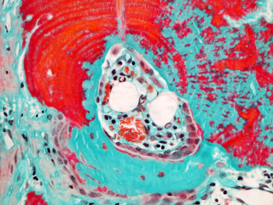
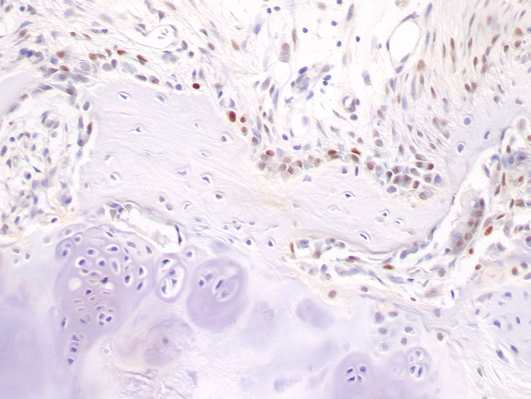
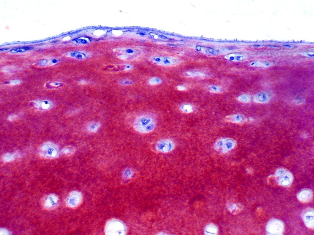
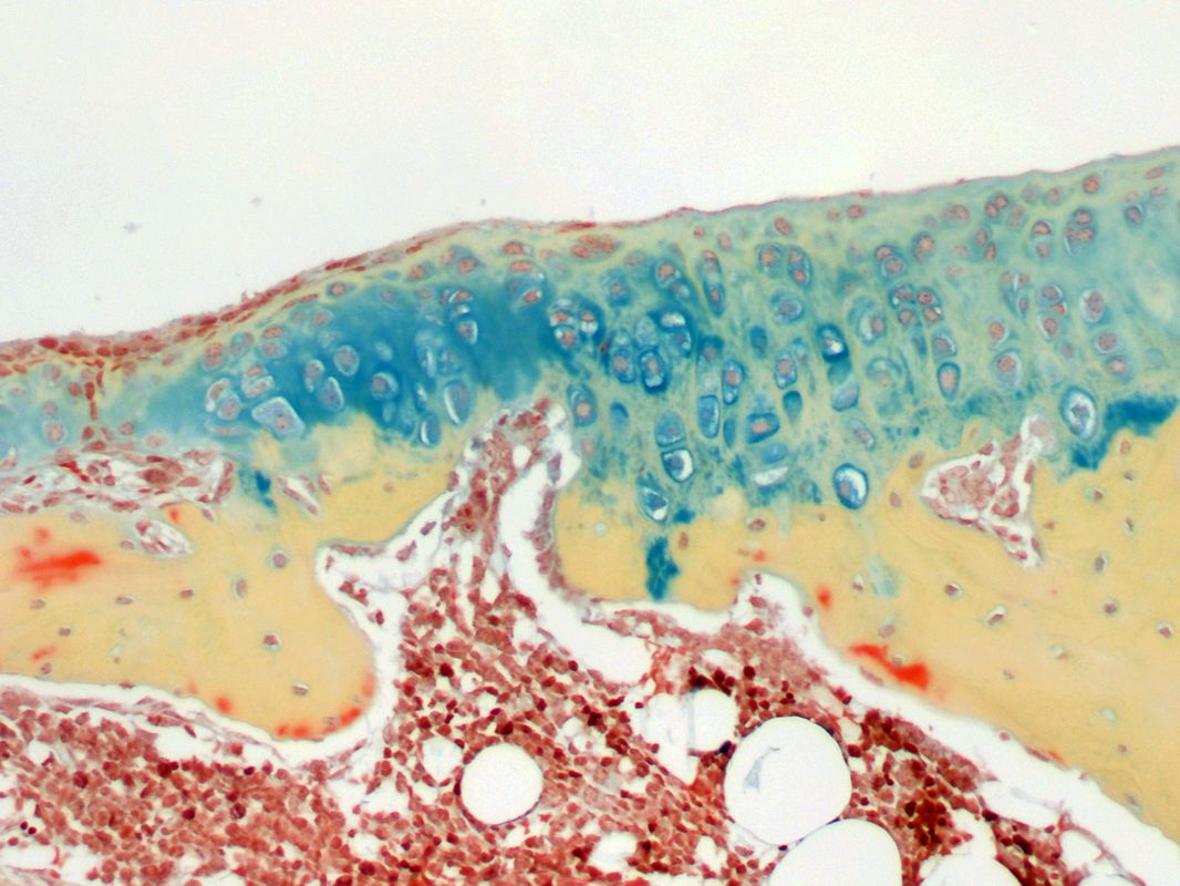
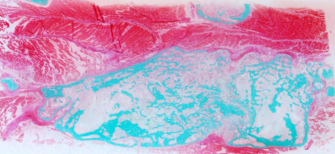
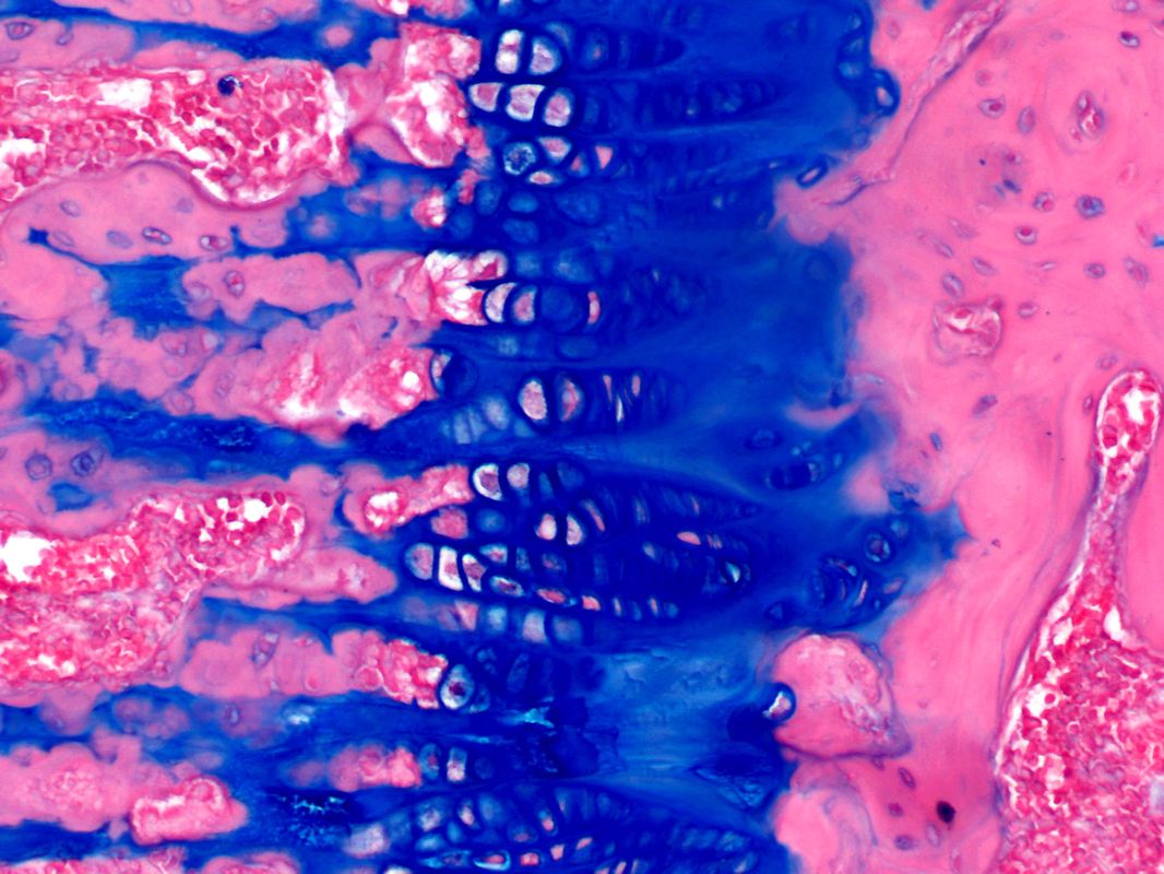 Growth plate; Alcian blue/H&E stain
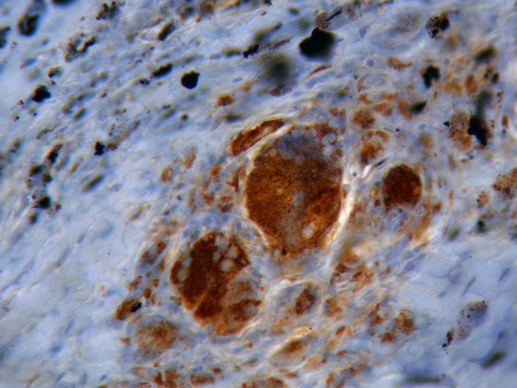
 Bone; Tartrate resistant acid phosphatase stain
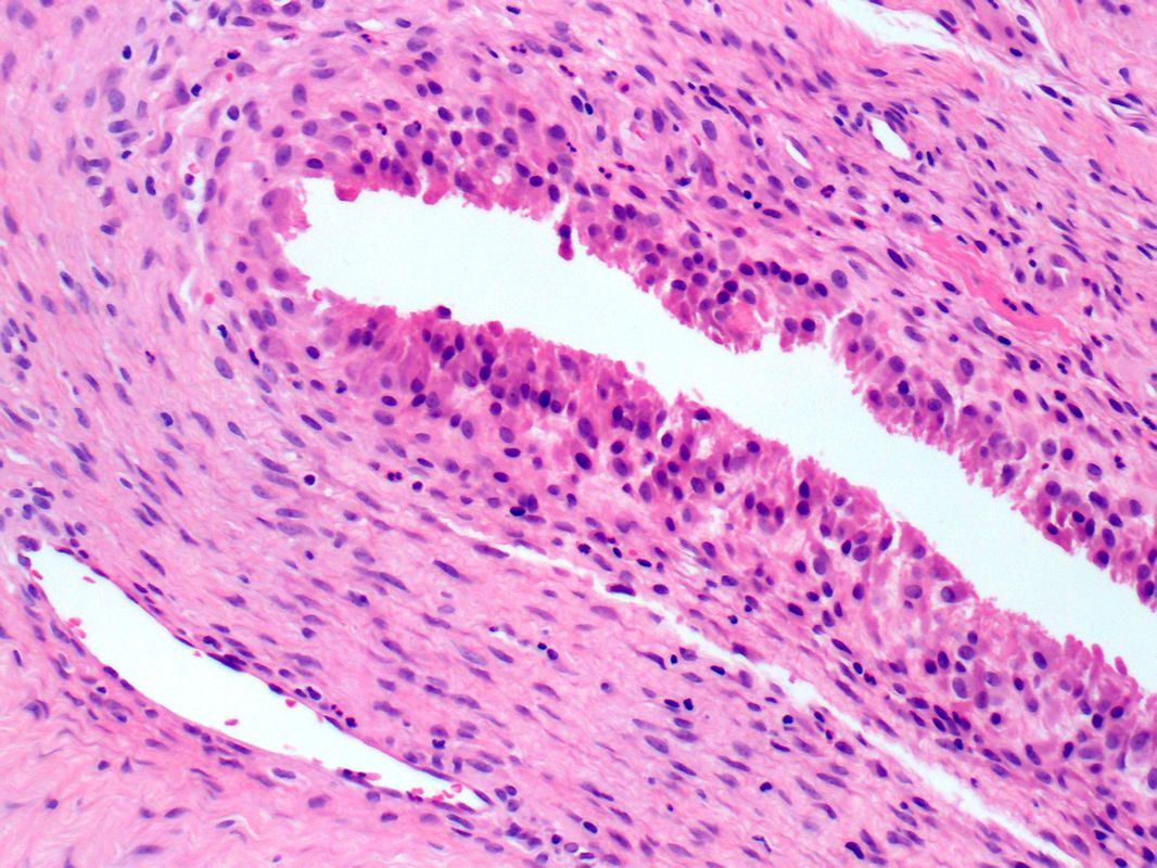 Synovium, hyperplasia; H&E stain
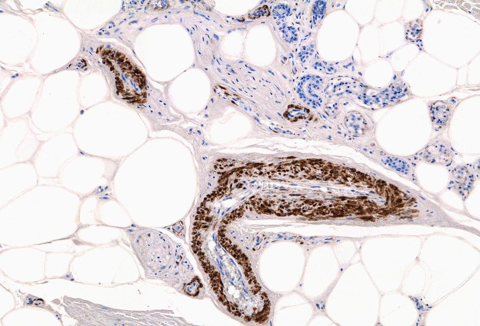
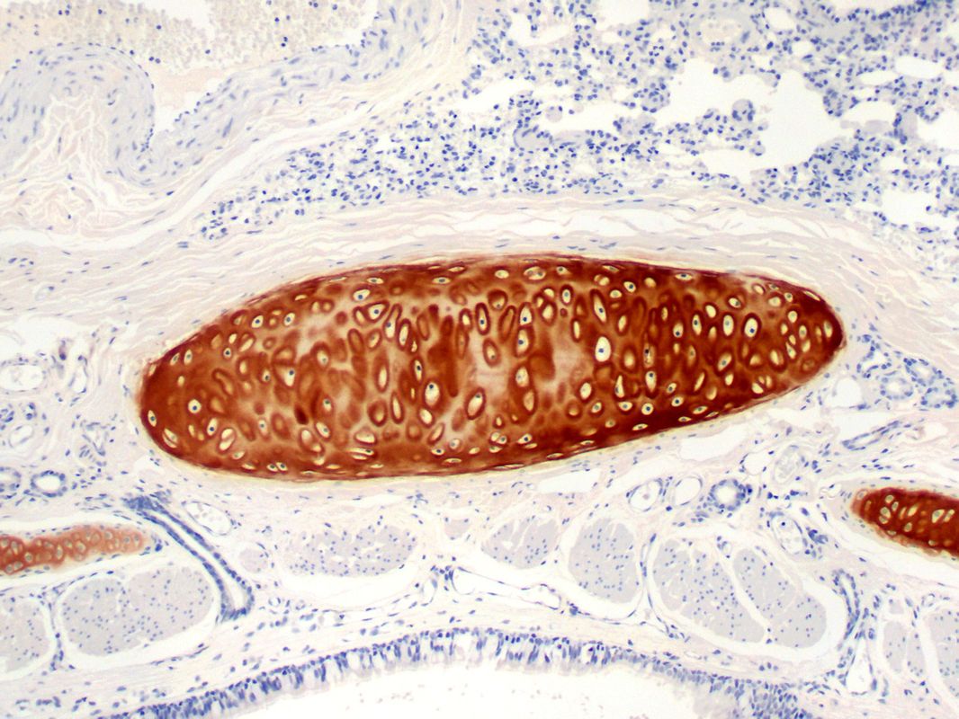 Trachea; Type II collagen IHC label
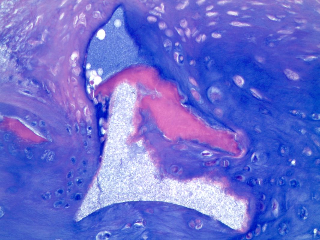
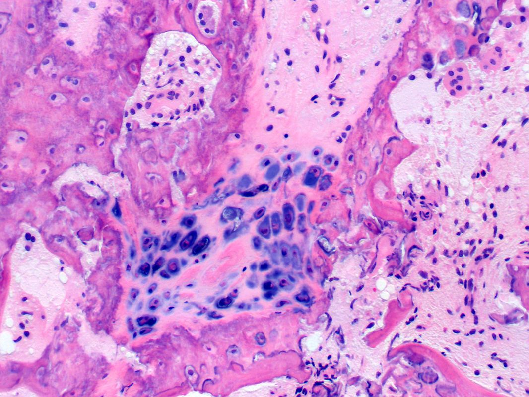
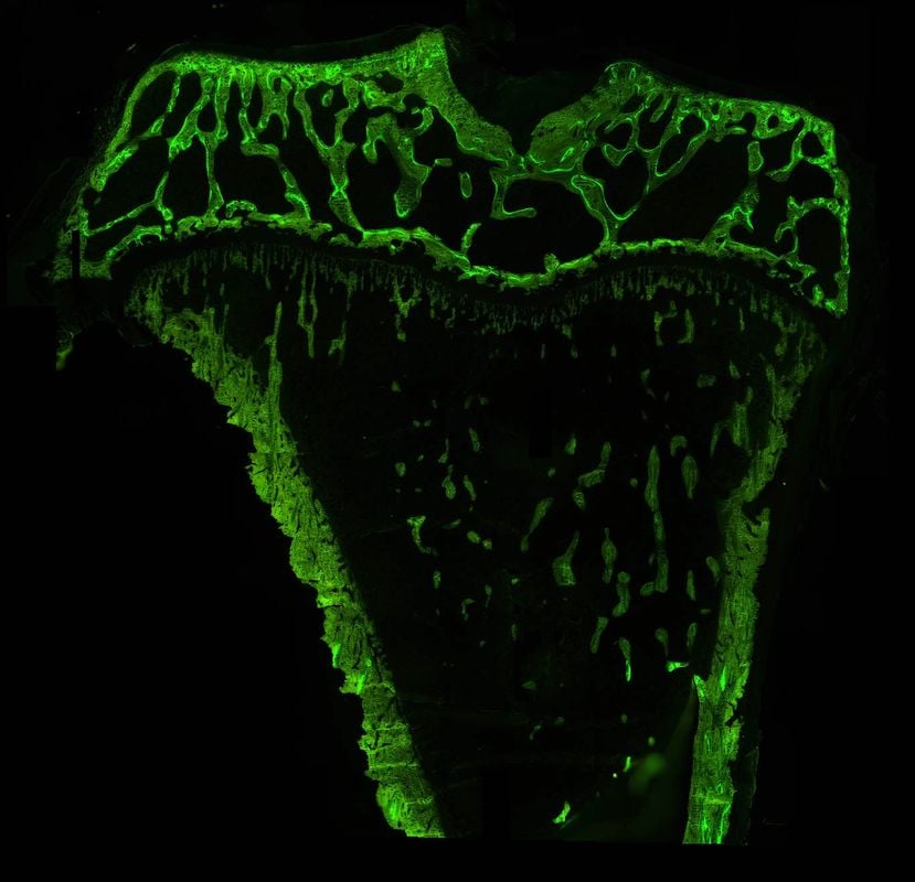
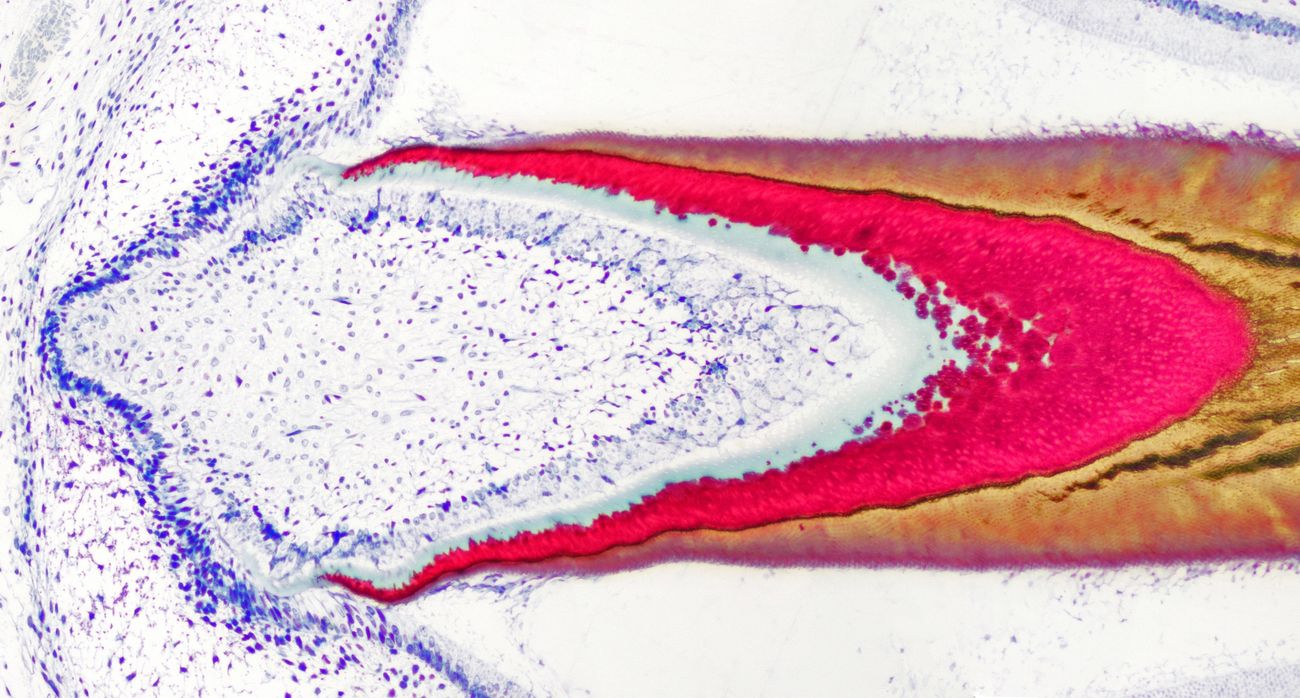 Tooth formation; Stevenel’s blue/van Gieson stain
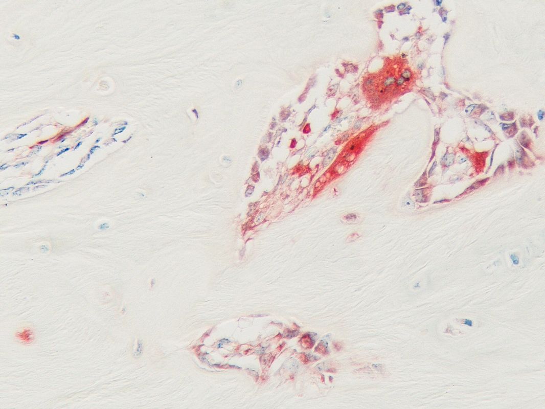
 Cartilage and synovium; Type II collagen, lubricin IHC double label
 Synovium-like tissue; Polymer implant particles (bright blue); Modified alcian blue stain
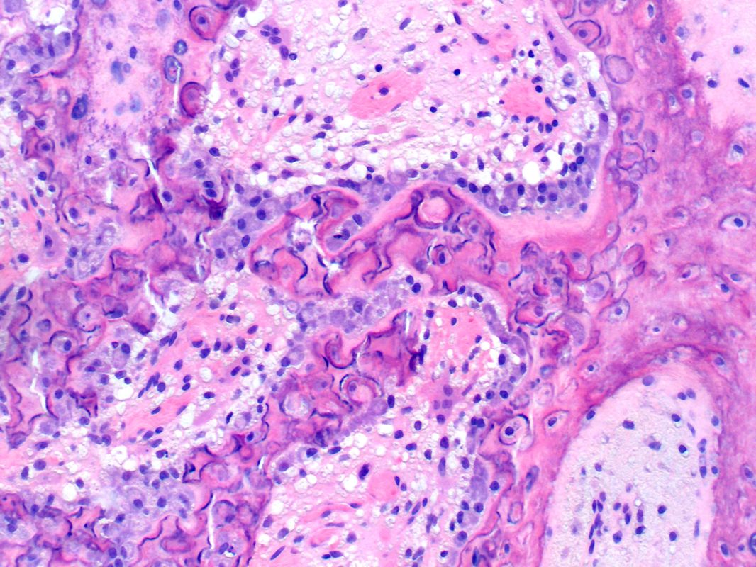 Endochondral bone formation; H&E
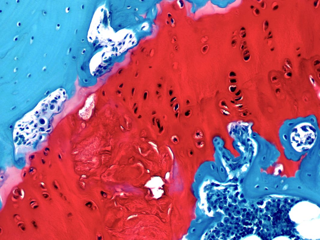
 Bone; Goldner’s trichrome stain
 Connective tissue; Smooth muscle actin IHC label
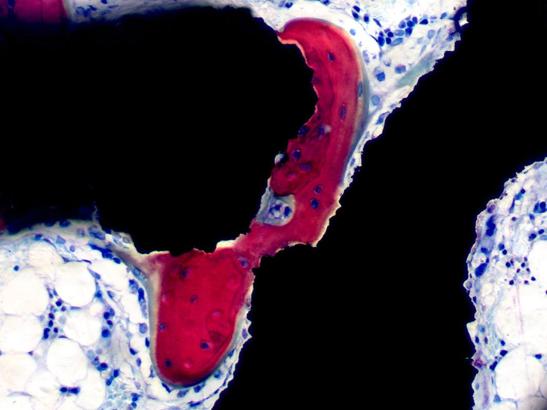
 Tibiofibular bone junction; H&E stain
 Growth plate; Safranin O stain
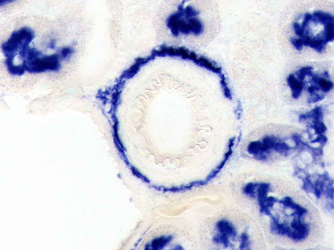
 Cartilage; Lubricin IHC label
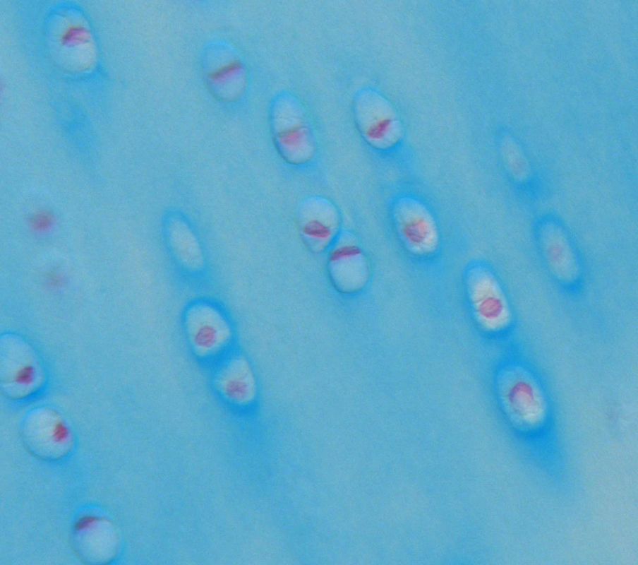
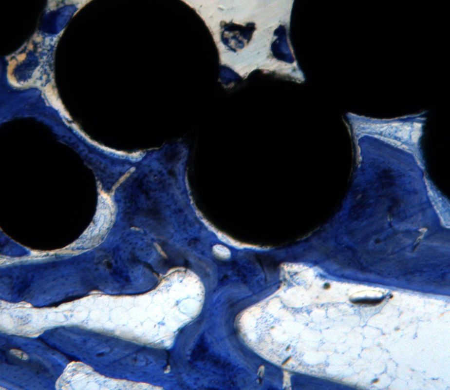
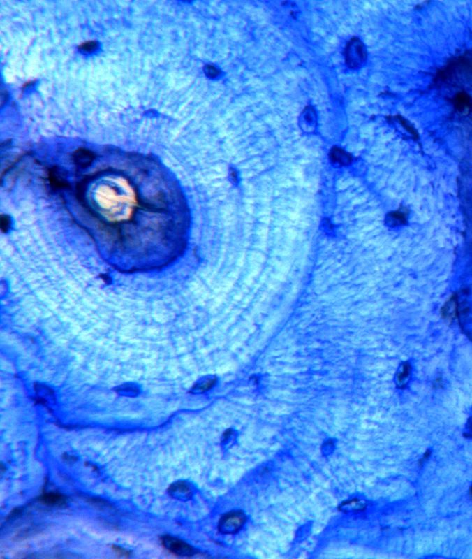 Cortical bone; Toluidine blue stain
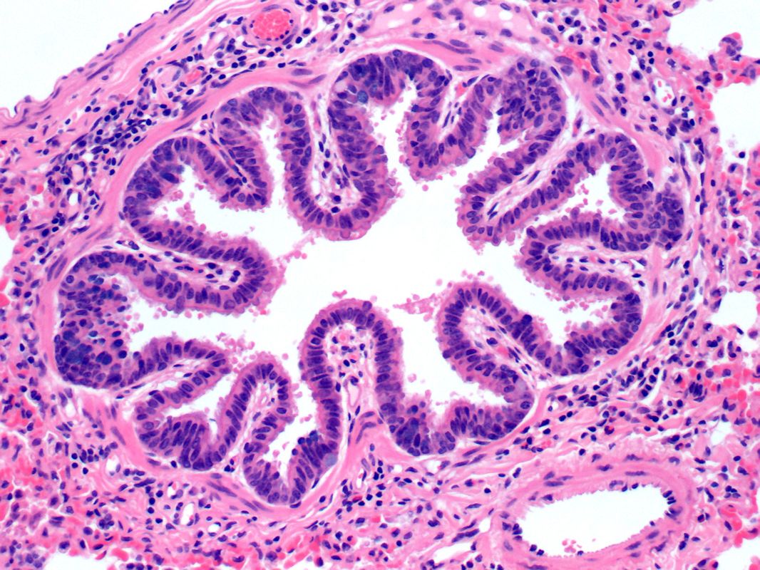 Bronchus; H&E stain
 Bone formation; Ceramic implant; Stevenel’s blue/van Gieson stain
 DBM bone induction; Goldner’s trichrome stain
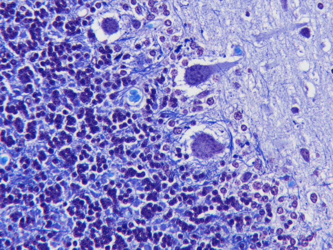 Cerebellum; Luxol fast blue, crystal violet stain
 Endochonral bone formation; H&E stain
 Herniated disc; PCNA IHC label
 Osteoclast/Osteoblast; Alkaline phosphatase (black)/Tartrate resistant acid phosphatase stain (red)
 Cartilage; Type II collagen and lubricin IHC double label
 Kidney; Alkaline phosphatase stain
 Bone and implant; Stevenel’s blue/van Gieson stain
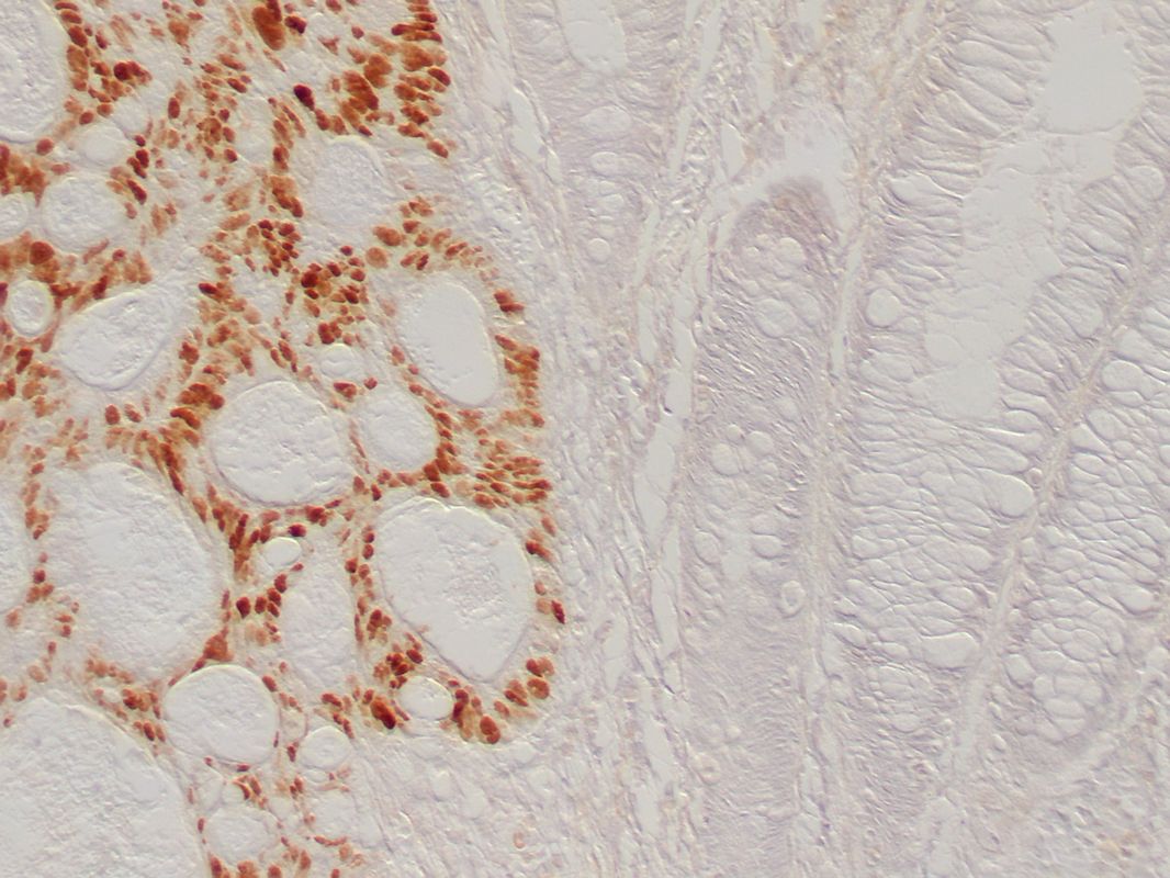
 Peri-metal implant tissue; Macrophage (CD68) IHC label; Ground section
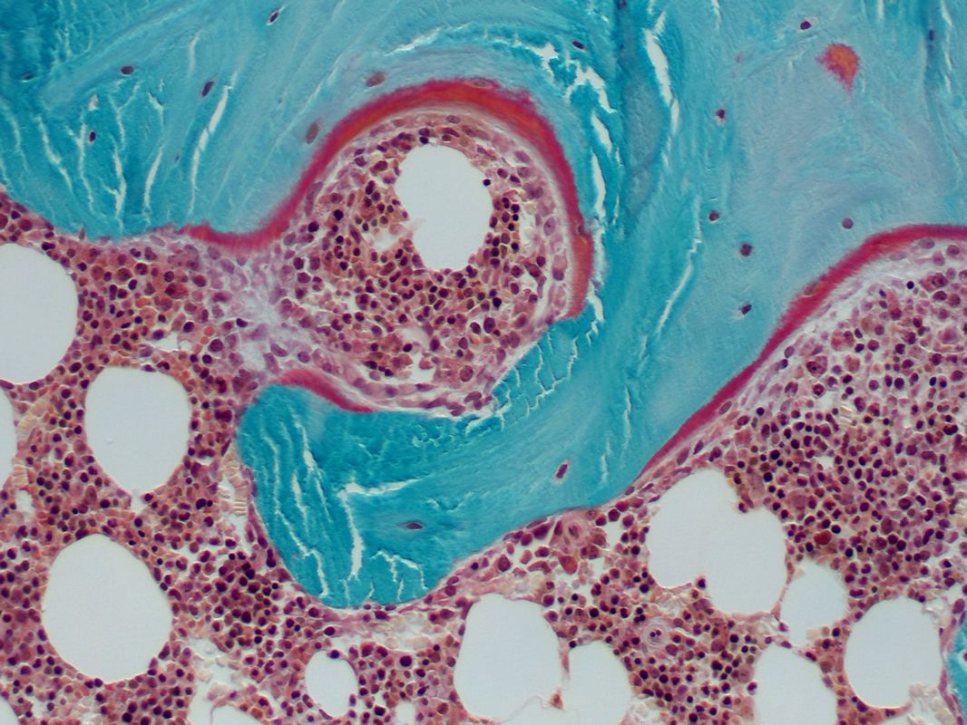
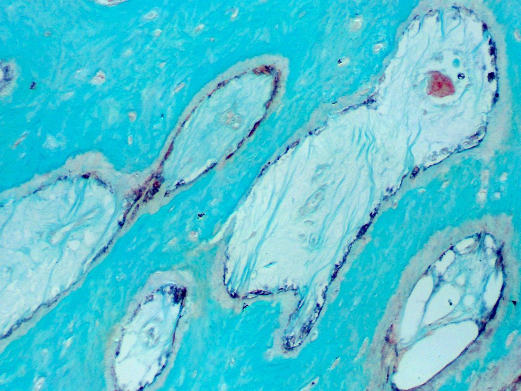
 Cartilage/bone; Movat’s pentachrome stain
 Bone cutting cone; Toluidine blue stain
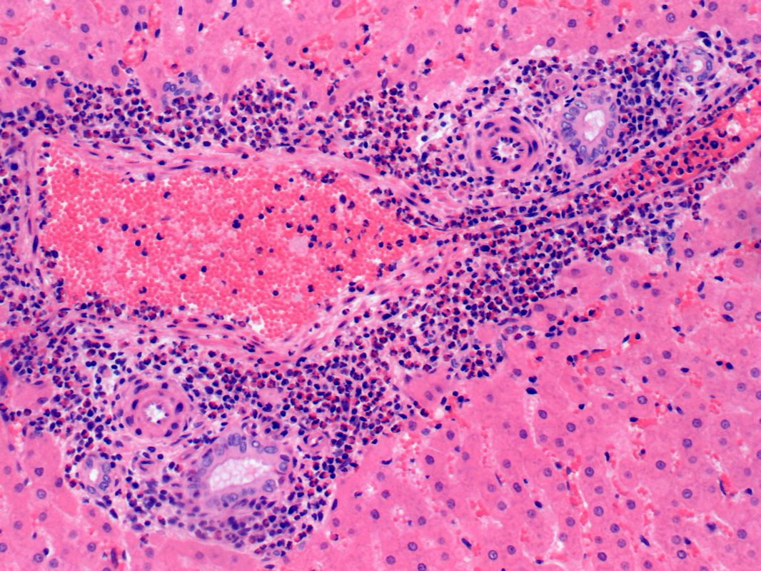 Liver; Inflammation; H&E stain
 Cartilage; Alcian blue stain
 Tibia; Tetracyline label
 Growth plate; Safranin O stain
 Bowel cancer; p53 IHC label
 Bone and implant; Toluidine blue stain
 Spine fusion; Goldner’s trichrome stain
Retina; H&E stain

Subcutaneous tissue; Metal implant; Toluidine blue stain

Skin; Picrosirius red stain

Growth plate; Alcian blue/H&E stain

Bone; Tartrate resistant acid phosphatase stain

Synovium, hyperplasia; H&E stain

Trachea; Type II collagen IHC label

Tooth formation; Stevenel’s blue/van Gieson stain

Cartilage and synovium; Type II collagen, lubricin IHC double label

Synovium-like tissue; Polymer implant particles (bright blue); Modified alcian blue stain

Endochondral bone formation; H&E

Bone; Goldner’s trichrome stain

Connective tissue; Smooth muscle actin IHC label

Tibiofibular bone junction; H&E stain

Growth plate; Safranin O stain

Cartilage; Lubricin IHC label

Cortical bone; Toluidine blue stain

Bronchus; H&E stain

Bone formation; Ceramic implant; Stevenel’s blue/van Gieson stain

DBM bone induction; Goldner’s trichrome stain

Cerebellum; Luxol fast blue, crystal violet stain

Endochonral bone formation; H&E stain

Herniated disc; PCNA IHC label

Osteoclast/Osteoblast; Alkaline phosphatase (black)/Tartrate resistant acid phosphatase stain (red)

Cartilage; Type II collagen and lubricin IHC double label

Kidney; Alkaline phosphatase stain

Bone and implant; Stevenel’s blue/van Gieson stain

Peri-metal implant tissue; Macrophage (CD68) IHC label; Ground section

Cartilage/bone; Movat’s pentachrome stain

Bone cutting cone; Toluidine blue stain

Liver; Inflammation; H&E stain

Cartilage; Alcian blue stain

Tibia; Tetracyline label

Growth plate; Safranin O stain

Bowel cancer; p53 IHC label

Bone and implant; Toluidine blue stain

Spine fusion; Goldner’s trichrome stain

